Thick Skin Histology Drawing
Histology integumentary 收藏 Histology dermis epithelial sebaceous physiology glands membrane corpuscles appendages krause receptors zapisano Skin histology anatomy hair scalp dermis follicle human diagram follicles microscope slide tissue layers wound healing glands thick microscopic structures
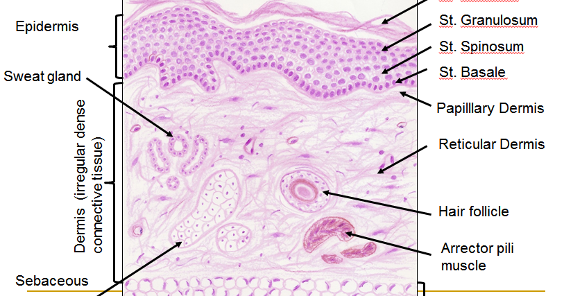
Histology Drawings: Skin (Integumentary System)
Layer hematoxylin histology integumentary epidermis eosin trichrome Histology integumentary anatomy stain tissue types layers mallory trichrome cutis Skin reading.php lab
Human structure virtual microscopy
Dermpath made simpleHistology of skin Histology drawings: skin (integumentary system)Scalp (skin).
Skin epidermis layers histology labIllustrations: thick skin Histology (skin)Stratum skin foot human thick histology corneum callus lucidum slide layers plantar integument spinosum basale granulosum pressure epidermis slides formation.

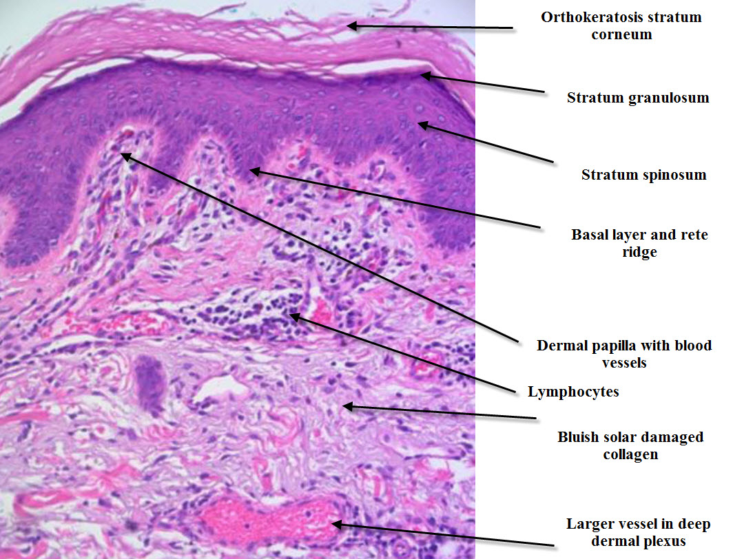
Skin (integumentary system)
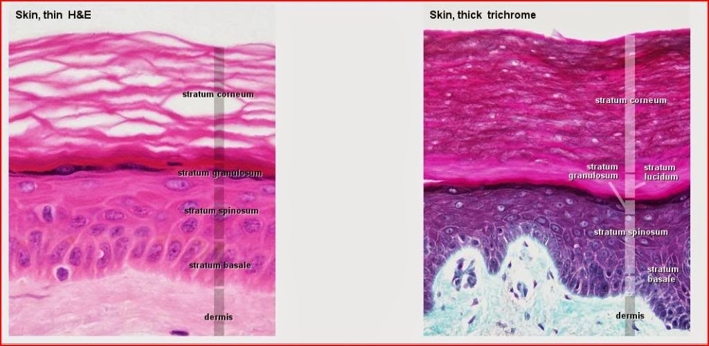
Skin thin thick histology microscope drawings between integumentary system light differences specimensSkin (integumentary system) Human histology soles microscopy palms friction fingertips specializedSkin histopathology dermatopathology simple introduction made inflammatory neoplastic dermpath email.
Histology drawings: january 2014Histology skin thin system integumentary drawings human anatomy thick section cross mallory trichrome slides 40x nervous cutis renal between .